
Lifeslittlesteps.com is a participant in the Amazon Services LLC Associates Program. As an Amazon Associate, I earn from qualifying purchases. Read full Disclosure here.
The navicular bone is a “boat-shaped” bone that is located in the midfoot. Navicular fractures are rare. Acute navicular fractures can occur during high-impact activities. Navicular stress fractures can also be seen in the foot due to repetitive activity. Navicular stress fractures represent 25% of all stress fractures.
When you sustain a navicular fracture, it’s important to be evaluated to determine the extent of the injury. This will determine the treatment that you will receive.
In this article, we will discuss navicular fractures of the foot in detail, and what you can expect regarding the treatment of these fractures.
Let’s dive in…
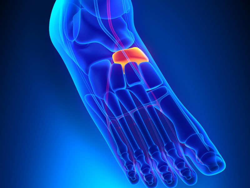
What Is the Navicular Bone in the Foot?
The navicular bone is one of the important bones that make up the midfoot. The navicular bone connects your ankle bone (talus) to the rest of the midfoot bones (cuneiform and cuboid bones).
Ligaments are also present that connect the navicular to adjacent bones. In addition, the strong posterior tibial tendon that is responsible for holding up the foot and supporting the arch inserts at the navicular bone.
The navicular bone receives its blood supply from the anterior and posterior tibial arteries. However, the blood supply is limited in the central portion of the navicular bone. This can make fractures that occur in this area at risk of not healing.
It’s important to understand the anatomy of the navicular bone, as this becomes important when it comes to the management of these fractures.
How Does a Navicular Bone Fracture Happen?
Acute navicular fractures can occur due to high-impact injuries to the foot. These include crush injuries, falls from heights, and motor vehicle accidents. In many cases, a part of the navicular may be fractured due to ligament avulsion that occurs during the injury. This is a “navicular avulsion fracture.”
Navicular fractures can also occur at the navicular tuberosity or the navicular body. Tuberosity fractures, body fractures, and avulsion fractures can be non-displaced (broken but in proper alignment) or displaced (fracture fragments are shifted).
During high-impact injuries, it’s not uncommon to see comminuted fracture fragments (multiple fragments). In addition, dislocation of the navicular may occur.
Stress fractures of the navicular bone can occur due to repetitive activity and pressure in the navicular bone. This can occur during sporting activities, prolonged marching, and even dancing. Navicular stress fractures can be non-displaced or displaced.

What Does a Broken Navicular Bone Feel Like?
- Pain and swelling on the inside of the foot and top of the foot
- Radiating pain that extends into the ankle
- Bruising of the midfoot
- Inability to bear weight on the foot due to pain
- Pain that worsens with activity but does not improve with rest
- Pain with walking during push-off
- Vague pain that worsens with time (in the case of a stress fracture)
How Do You Treat a Fractured Navicular?
If you suspect you have a navicular bone fracture, make sure to go to the Emergency Room or see your foot doctor.
Your doctor will perform a physical exam. Your doctor will check your pulse to make sure your blood flow to the foot is good. Your doctor will also check your skin integrity to make sure there are no lacerations in the skin.
X-ray
Your doctor will order x-rays to evaluate the foot. He/she will check to see where the fracture is in the navicular bone, and if it is non-displaced or displaced. Your doctor will check to see if the navicular is dislocated. Your doctor will also check to see if the navicular fracture is comminuted (fracture has greater than two pieces).
CT scan
Midfoot fractures can sometimes be difficult to see on the x-ray. Your doctor may order a Computed Tomography scan (CT) which will show fracture lines in more detail.
MRI scan
Your doctor may choose to order a Magnetic Resonance Imaging Test (MRI) which can show the fracture line as well as identify bone marrow edema that may be visible during a stress fracture. An MRI will also identify any tendon and ligament injuries.
Once all this information is obtained, appropriate treatment for the fracture will be determined.
Read more: Navicular Fractures: How To Diagnose This Injury
Non-displaced Fractures of the Navicular
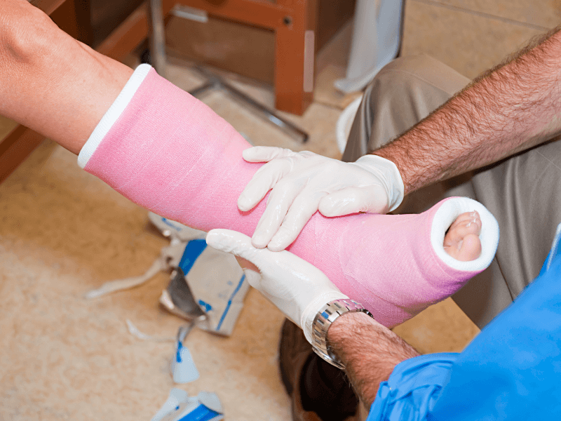
If the navicular in your foot is broken, you should stay off of your foot for 6-8 weeks minimum in a cast or a walking boot.
Fracture healing takes 6 to 8 weeks minimum. By walking prematurely on a broken bone, you can displace the fracture and cause further injury.
Your doctor will order serial x-rays every 3-4 weeks to assess the fracture. If the fracture is healed after 6-8 weeks, you can slowly transition to weight bearing in the boot for another 3-4 weeks before transitioning into athletic shoes.
Displaced Navicular Fractures
If the navicular fracture in your foot is displaced, your doctor will suggest surgery. Fractures that show greater than 3mm of displacement will need surgery. In addition, if there is greater than 1mm of joint displacement, your doctor will recommend surgery.
During high-impact injuries, the navicular bone can become crushed and no longer articulate with the joints properly. This can cause chronic pain if it is not fixed.
Your doctor will perform surgery in the operating room under anesthesia. He/she will repair the fracture using bone plates/screws. Your doctor may also insert a bone graft into the navicular to fill a defect in the fracture site.
You will need to stay non-weight bearing in a cast or cast boot for up to 8 weeks after surgery. Your doctor will order x-rays every 3-4 weeks to assess healing. After the bone is healed, you may need rehabilitation to help you walk again. This is important to regain strength in the foot.
It could take up to 3 months before you are walking comfortably in athletic shoes.
Open Fractures of the Navicular
If you have a large laceration with a fracture of the navicular bone, you will need to be admitted to the hospital for IV antibiotics. Your doctor will check if there is any vascular injury and consult a vascular surgeon for repair if blood supply is compromised.
If not, your doctor will perform surgery to clean the open fracture site and stabilize the fractured navicular using fixation.
You will need to remain off your foot for a minimum of 8-12 weeks in a cast or splint.
Delayed Healing of the Navicular Fractures
At times, even when surgery is performed, healing of navicular fractures can be delayed. This is due in most cases to reduced blood supply in the central portion of the navicular.
In addition, people who smoke, have diabetes, and have low calcium/Vitamin D levels are at risk of not healing. A “non-union” can form. This can be painful.
If this is the case, your doctor may suggest using an external bone stimulator.
An external bone stimulator is a machine you wear for 20 minutes a day. Depending on the machine used, ultrasonic or electrical stimulation is used to stimulate fracture healing. Your doctor may recommend you wear the bone stimulator for 3 months or longer.
If you smoke, you should try to quit smoking, as smoking can impact bone and wound healing.
Diabetics should focus on blood sugar management, as elevated blood sugar levels can slow wound healing.

Many people have low Vitamin D levels. Low levels of calcium and Vitamin D can impact bone healing. If you are deficient, your doctor may recommend a calcium and Vitamin D supplement in addition to diet modifications.
Orthotics for Navicular Pain
After your navicular fracture is healed, you should consider wearing orthotics. Orthotics help to lift the arch of the foot and thus alleviate pressure from the navicular. This is important because it can help prevent repetitive stress fractures. Orthotics help to rebalance forces throughout the foot.
Custom orthotics are ideal, because they are custom-made to fit your feet. Ask your foot doctor for a prescription for custom orthotics.
Conclusion
In conclusion, navicular fractures are serious and should be addressed quickly to optimize good outcomes. As mentioned in this article, treatment for navicular fractures will vary depending on the extent of the injury. If you suspect that you have a navicular fracture, make sure to contact your foot doctor immediately.
Related article: Pediatric Navicular Fractures- How To Diagnose and Treat Them
SHARE THIS PIN!
References
- Shakked RJ, Walters EE, O’Malley MJ. Tarsal navicular stress fractures. Curr Rev Musculoskelet Med. 2017 Mar;10(1):122-130. https://www.ncbi.nlm.nih.gov/pmc/articles/PMC5344863/
- Sanders, Roy MD*,†; Serrano, Rafael MD†. Navicular Body Fractures—Surgical Treatment and Radiographic Results. Journal of Orthopaedic Trauma: February 2020 – Volume 34 – Issue – p S38-S44. https://journals.lww.com/jorthotrauma/fulltext/2020/02001/navicular_body_fractures_surgical_treatment_and.9.aspx.
- Kim WJ, Jung KJ, Yeo ED, Lee HS, Won SH, Lee DW, Ji JY, Yoon SJ, Hong YC. Treatment of Navicular Stress Fracture Accompanied by Os Supranaviculare: A Case Report. Medicina (Kaunas). 2021 Dec 24;58(1):27. https://pubmed.ncbi.nlm.nih.gov/35056335/
- Attia AK, Mahmoud K, Bariteau J, Labib SA, DiGiovanni CW, D’Hooghe P. Return to sport following navicular stress fracture: a systematic review and meta-analysis of three hundred and fifteen fractures. Int Orthop. 2021 Oct;45(10):2699-2710.https://link.springer.com/article/10.1007/s00264-021-05147-6
- Ostlie DK, Simons SM. Tarsal navicular stress fracture in a young athlete: case report with clinical, radiologic, and pathophysiologic correlations. J Am Board Fam Pract. 2001 Sep-Oct;14(5):381-5. https://pubmed.ncbi.nlm.nih.gov/11572544/
- Rosenbaum, A. J., Uhl, R. L., & DiPreta, J. A. (2014). Acute fractures of the tarsal navicular. Orthopedics, 37(8), 541-546.https://journals.healio.com/doi/abs/10.3928/01477447-20140728-07
- TOWNE, L. C., BLAZINA, M. E., & COZEN, L. N. (1970). Fatigue fracture of the tarsal navicular. JBJS, 52(2), 376-378.https://www.aafp.org/pubs/afp/issues/2003/0101/p85.html
DISCLAIMER: The information, including but not limited to, text, graphics, images and other material contained on this website are for informational purposes only. No material on this site is intended to be a substitute for professional medical advice, diagnosis, or treatment. Always seek the advice of your physician or another qualified healthcare provider with any questions you may have regarding a medical condition or treatment before undertaking a new healthcare regimen, and never disregard professional medical advice or delay in seeking it because of something you have read on this website.


One thought on “Navicular Fractures- What You Need to Know”
Comments are closed.